Printing human organs – is this the answer to solving the transplant shortage?

There are approximately 10,000 people on the UK transplant list waiting to receive an organ. Statistics show that, due to a shortage in organs available for transplant, every day 3 of these people will die before receiving their transplant. Unfortunately, we have not yet found a way to address this shortage. However, with this in mind, money is being poured into investigating new technologies aimed at tackling this problem.

But how? Well, the answer may have been under our noses all along. Using a popular everyday technology, we may be able to use printing with a modern twist to generate these desperately needed organs. Imagine being able to print out a new liver, or a lung, or in fact whatever organ you needed.

Kidney 1In 2011 Dr. Anthony Atala, director at the Wake Forest Institute for Regenerative Medicine, received a standing ovation when, on stage during a presentation, he printed out a prototype kidney made from living cells. Although far from perfect, the kidney was able to break down toxins and produce a waste-like product, just like the genuine thing. Despite their diminutive size, these ‘miracle organs’ have the potential to revolutionise medicine as we know it.

And these aren’t the only organs to have been produced using 3D printing. Scientists at Organovo, a bioprinting specialist, have now created miniature livers. These tiny livers are just 4mm wide and ½ mm in depth, but are still able to produce the proteins essential for carrying hormones and drugs. Importantly, the livers produce cytochrome p450, which is vital for breaking down drugs in the body. What’s more, cells from blood vessels can be incorporated into the livers to supply them with oxygen and other nutrients.

printer 13D printers work by printing out layer upon layer of ink (or any other substance) until a 3D object is achieved.   All we have to do is tell the printer what to print with a computer aided design program, hit the print button and let the magic commence. Hey presto- there you have a real-life 3D object.

When printing organs, layers of living cells are laid out in sheets upon each other and a scaffolding material called hydrogel is spread between the layers providing nutrition to the cells and helping them fuse together. The cells are able to survive for as long as four months in these conditions. Images of the patient’s original organs are generated using a CT scan which forms a template that instructs the printer on how to to produce a life-like replica of the organ.

Although 3D printing may seem ultramodern, it has actually been used in the medical field some time to produce things such as hearing aids and braces.  This technology has astonishingly also been used to create an entire lower jaw for an elderly women who was deemed too fragile to undergo reconstructive surgery. The jaw was designed to be realistic, with cavities and grooves in the mould to allow for the re-attachment of jaw muscles and nerves.

We are now at a stage where we are able to print sheets of living cells, but only on a small scale. Researchers have surmounted a massive hill, and have finally found a way to pass human embryonic stem cells through a 3D printer without sustaining any significant damage to the cells.

Researchers are hopeful that over the next few years we will be able to use these tissue strips to test the toxicity of new drugs before exposing living patients to these potentially dangerous substances, saving not only time and money but also reducing risks to patients. Using the so-called mini organs, researchers will be able to watch the progression of diseases in life-like organs outside of the body.

Over the course of the next 10 years we will most likely see 3D printing being used to assist in regeneration such as bone and skin grafts, patches for heart conditions, regeneration of sections of blood vessels and replacement cartilage for joints.

As for producing entire organs, there is still a long way left to go. The problem lies with being able to produce a fully functioning organ on a full-size scale with the ability to maintain its own oxygen supply via a vascular system, and to remove its own waste.           The hope is that researchers will be able to devise a way that will allow a printer to produce a full scale organ without any damage to the cells, and that is sufficiently supplied with oxygen and nutrients. If we manage to achieve this, then using ‘printed’ organs may cease to be something of a dream and become a reality. Over the next 10 or 20 years, the thousands of people likely to need a transplant could be lucky enough to receive an organ almost immediately instead of waiting months or even years as is the case now.

Post by: Sam Lawrence

For more information on organ donation or to join the organ donor register visit: http://www.organdonation.nhs.uk/

The Komodo dragon: how this foul-mouthed fiend feasts

“Meddle not in the affairs of dragons, for you art crunchy and good with ketchup.” ― Anon

Komodo1
A wild Komodo dragon

The fearsome Komodo dragon (Varanus komodoensis) is the world’s largest living lizard, weighing in at an average of 150 lbs, and measuring 3 metres in length. It is native only to five of Indonesia’s islands, including its namesake, Komodo.

The Dragon is both a scavenger and an ambush predator; capable of lying in wait for hours at a time until prey, such as buffalo, come close enough for it to attack. Of course, not all kills are clean and quick. Animals injured in attacks by Komodo dragons can survive only to die days or weeks later from their wounds. Dragons have even been known to follow wounded prey for days until they die in order to enjoy an easy meal.

Interestingly, being the dominant predator in their habitat, it is likely that a member of the species will benefit from a delayed kill; even if it isn’t the individual that caused the death. This is very possibly a demonstration of ‘altruistic’ behaviour whereby an individual’s actions benefit the population as a whole.

But what exactly do these injured animals die from? Certainly Komodo dragon bites can be sufficiently deep to cause fatal blood loss but this is not always the case. Something else must come into play. The nature of that ‘something’ has caused a lot of controversy in recent years as different studies have produced contradictory results. There are currently two major theories, the oldest of which is coming under increasingly heavy fire from supporters of its more contemporary rival:

The Original Theory: Harmful bacteria live in the mouth of the Komodo dragon and infect wounds the dragon inflicts upon its prey

This particular theory originated in Walter Auffenberg‘s book, ‘The Behavioral Ecology of the Komodo Monitor’, published in 1981. In order to understand why Dragon bites sometimes resulted in delayed fatalities, Auffenberg swabbed the gums of captured Komodo dragons and identified four bacterial species from the samples. Three were described as being common causes of infections in animal bites.

Around 20 years later, Joel Montgomery’s group repeated this test, but on a much larger scale and with far more sophisticated screening techniques. They identified 58 species, with considerably more seen in wild Dragons than captive ones. The group also injected Dragon saliva directly into the abdomens of mice in the hope that they might recover a bacterial culprit from any mice that died from these simulated ‘bites’.

Some, however, consider their results contentious. All of the bacterial species the group found are pretty common in soil, plants or on animals’ skin. One species – Pasteurella multocida – present in some of the infected mice, was assumed to play an important role in prey infection, since the Dragons themselves were immune to it. Unfortunately, the species was only present in 5% of Dragons’ mouths and it turns out it isn’t actually capable of causing fatal septicaemia at the rate seen in Dragons’ prey.

Komodo2
Young Komodo Dragon feeding at a water buffalo corpse on Rinca

 It is now increasingly thought that the bacteria found in Komodo dragons’ mouths are simply the bacteria that were growing on/in the reptiles’ most recent meals. This may explain why captive Dragons, which are fed fresh meat, have fewer bacterial species in their mouths than their wild relatives, which will eat rotting carcasses. This all makes for a conspicuous lack of compelling evidence to support the idea that Dragons have evolved to use bacteria as a ‘weapon’.

The Modern Theory: The Komodo dragon uses venom to incapacitate its prey

This idea arose from a 2009 study by Bryan Fry’s group in Australia. They noticed that prey wounded by bites from Komodo dragons show common symptoms; namely being subdued, bleeding heavily and finally going into shock. Interestingly, these are the same symptoms caused by venom released by members of a related genus of lizards called the Helodermatids.

With this in mind, Fry’s group performed a Magnetic Resonance Imaging (MRI) scan of a preserved Dragon’s head and discovered a large venom gland in the lower jaw. The gland was divided into 6 compartments, with separate ducts leading from each compartment into the mouth, opening out between the Dragon’s serrated teeth.

The team discovered that the venom contained 2,000 proteins, around a third of which were known toxins in related reptile species. Indeed, the Dragon’s venom composition was similar to that of snakes, containing anticoagulants and toxins that lower blood pressure, causing persistent bleeding and weakness.

Interestingly, unlike other venomous lizards, Dragons do not have teeth specially adapted for chewing and working venom into their unlucky victims’ wounds. Instead, Fry suggests that the Dragons, who don’t have particularly strong bites, use their serrated teeth to “grip-and-rip” flesh, forming deep wounds into which venom could easily flow.

Continuing controversy:

The venom theory has more convincing evidence behind it than its predecessor and has become a far more widely accepted theory since its conception in 2009. It seems far more feasible that Dragons could have evolved effective venom, rather than evolving a co-dependent relationship with a variable bacterial population. The next steps in proving this theory may be to find evidence of Dragon venom in a prey animal, as well as physical proof that these venom glands actively work.

Despite all of this, however, the original theory still has a lot of supporters and has not yet been entirely ruled out. It is likely that the scientific community will remain split on the issue for some time to come. One thing we can all agree on, though, is that this fascinating beast has evolved a highly effective killing mechanism, which makes it both a powerful and intimidating predator.

Guest post by Ian Wilson

Learn a little more about Ian:

IanWilson“I’m currently about to enter my fourth and final year of a PhD studying the genetics of the human parasite Entamoeba histolytica. I’m based at the University of Liverpool, which is also where I completed my undergraduate degree in Microbiology. I’m really passionate about improving the public’s relationship with science and I aim to become a full-time science communicator when I finish my PhD so I can really get stuck in!”

You can follow Ian on twitter @Science_Gremlin and for more top-notch science writing, including an answer to the question: Just how scientifically possible are Gremlins?: Visit his blog: www.sciencegremlin.wordpress.com

Science of the bloody brain

mosso
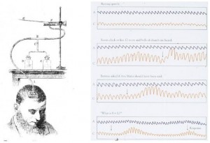
One of Mosso’s experiments. Each of the four traces on the right compares brain blood flow (red) and pulsations in the feet (black) simultaneously, during 1)resting 2)listening to the clock and church bells 3)remembering whether Ave Maria should have been said and 4)’8×12′?

Luigi Cane literally had a hole in his head. A brick had unforgivingly fallen on the back of it, smashing a section of his skull like a spoon knocking the shell off the top of a hard-boiled egg. And so, after surgery, part of the surface of his brain was left precariously unprotected except for a layer of skin. Peering through this accidental window into his head, Dr. Angelo Mosso was able to measure the pulsations of the brain’s blood supply. Cane sat in Mosso’s lab with pressure gauges strapped around his feet and a handmade instrument resting delicately on the skin over his vulnerable brain. This was to be the world première of neuroimaging.

Angelo Mosso, a 19th century physiologist and first brain imager

“What is 27 times 13?” Mosso inquired. Cane thought deeply and silently while the various contraptions simultaneously showed his feet shrinking while his brain swelled with blood flow. This experiment was the first to reveal that when our mental ‘cogs’ turn, a boost of blood is directed to the brain. Mosso confirmed this in individuals with intact skulls with what was essentially a wobble-board bed. When people lying down on the balance thought about tricky or even particularly emotional questions, it would tip down towards the head end with the weight of the extra blood.

The brain is an extremely greedy part of the body when it comes to blood. While it only makes up about a fiftieth of the body’s mass, it consumes up to a fifth of the total energy and oxygen carried in the bloodstream. Charles Roy and Charles Sherrington later proved that the blood rushing to the head was actually being diverted specifically to the parts that were most active – like a bonus for the busiest brain cells. Over twelve decades later, neuroscientists are still using this same principle to observe brain activity and the accompanying ‘rush of blood’ to the head.

The brain imaging technique functional magnetic resonance imaging (fMRI) works on the principal that deoxygenated haemoglobin (the protein that carries oxygen in red blood cells) has magnetic properties. In essence, fMRI can measure how well-oxygenated or deoxygenated different parts of the brain get when the person in the scanner performs a task, for example reading, writing, or thinking about chocolate. But information collected from this kind of experiment needs to be handled very carefully.

Computer-enhanced fMRI scan of a person who has been asked to look at faces. The image shows increased blood flow in the part of the visual cortex that recognizes faces.

Firstly, fMRI is not a direct measure of brain activity per se; rather, it’s the triggered oxygenated blood flow response to brain activity. Secondly, no one really knows what a larger blood flow response means, especially in parts of the brain that have several jobs. Lots of blood in a specific part of the brain while doing sums might mean that a person can do sums easily because their blood supply is so efficient. Alternatively, it could be interpreted as suggesting that person struggles with mental arithmetic and needs more blood in their head to cope. Thirdly, fMRI data needs to be stringently tested to avoid seeing activity that isn’t there. Researchers at the University of California found that using different statistical tests they could see a blood flow response in the brain of a dead salmon while it was looking at different human faces – and won an IgNobel Prize for highlighting the dangers of shoddy stats.

With all this to bear in mind, it’s perhaps unsurprising that poorly carried out fMRI experiments have been dubbed the modern phrenology – the practice of comparing measurements of peoples’ skulls to infer personality traits. What is perhaps more surprising, though, is that despite the speculations on the validity and accuracy of fMRI, it is being used for things besides its more traditional remit. ‘No Lie MRI’ is a company in the U.S. that advertises the use of brain imaging to detect liars or untrustworthy individuals, whether they be potential politicians, investments or romantic interests. Brain imaging techniques including fMRI have even controversially been used as evidence in Indian courts of law.

pain
Example of fMRI responses to painful heat to the forehead in a cohort of 12 subjects. ACC, anterior cingulate cortex; PCC, posterior cingulate cortex (Moulton et al., unpublished observations). Borsook et al. Molecular Pain 2007 3:25

There are, however, other emerging uses for fMRI that may improve its reputation. By watching live feedback of the blood flow going to the anterior cingulate and insula, two pain centres deep within the brain, sufferers of chronic pain can consciously train these parts of the brain to receive more blood. Christopher deCharms and his colleagues at Omneuron have found that people who were given the real, live feedback from their insula and cingulate and successfully learnt to train the blood flow within these parts said they experienced less pain than usual. Conversely, people unwittingly shown a dummy feedback (random fluctuations or blood flow levels from an unrelated part of the brain) didn’t report any substantial pain relief.

Brain imaging techniques that rely on measuring blood flow around the brain should be carefully interpreted; fMRI is heavily-used in research and is still fashionable in brain research. Technology has come on a massively long way since the days of wobble boards, so we should probably count ourselves lucky that we don’t need a hole in our heads to unlock the further mysteries of the blood in our brains.

Post by Natasha Bray


Hope for new MS drug which could repair damaged cells.

Researchers from the private biotech firm ENDECE Neural have just announced the development of a new compound they believe may have the potential to repair damage caused by multiple sclerosis (MS).

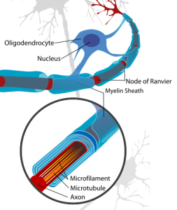
MS is the most common neurological disorder affecting young adults in the western hemisphere. Although scientists are still unsure of what causes the disorder, it is known that symptoms stem from damage to the fatty covering surrounding nerve cells, known as the myelin sheath. It is believed that in the early stages of the disease the body’s own immune cells (cells usually primed to seek out and destroy foreign agents within the body, such as viruses and parasites) mistake myelin for a foreign body and launch an attack. Since myelin is essential for fast neural communication and cell protection, the symptoms of MS stem from a slowing of neural communication and ultimately nerve cell damage.

oligoThe myelin surrounding cells in the brain and spinal cord is provided by cells called oligodendrocytes. These cells reach out a number of branching arms which wrap around segments of surrounding neurons, forming the myelin sheath. The majority of drugs available for treatment of MS aim to reduce initial damage to this sheath. However, researchers from ENDECE are now investigating treatments which can increase the number of oligodentrocytes in the central nervous system, thus leading to remyelination of damaged cells. Dr. James G. Yarger, CEO and co-founder of ENDECE notes, “For decades, researchers have been seeking ways to induce remyelination in diseases such as MS that are characterized by demyelination,”. And now this dream may be becoming a reality.

ENDECE’s work revolves around their pipeline drug NDC-1308. Although the name isn’t likely to turn any heads, its properties just might. Following the observation that pregnant women typically do not experience the symptoms of MS during their third trimester, a number of researchers have been exploring a possible role for estrogen in the treatment of MS. ENDECE researchers created 40 separate estradiol analogues (substances similar in structure to estradiol but with a range of key modifications) and assessed their biological effects. From this work they found that one analogue (NDC-1308) had a particularly potent effect on oligodentrocyte precursor cells (OPCs – cells with the ability to become mature oligodentrocytes), causing them to differentiate into mature oligodendrocytes. In follow-up studies researchers found that treatment with NDC-1308 led to remyelination in a mouse model of MS, specifically showing a 20% increase in myelination in the hippocampus (a region of the brain known to experience demyelination in this model). NDC-1308 was also found to cause remyelination in the rat and to induce cultured OPC cells to differentiate into mature oligodendrocytes. Taken together, these findings suggest that NDC-1308 may prove effective in restoring the lost myelin sheath on damaged axons in patients with MS.

Dr. Yarger states, “We envision NDC-1308 being administered either alone or in combination with current therapeutics that target the immune response and/or inflammation associated with MS. By inducing remyelination, it may be possible to restore muscle control, mobility, and cognition in patients with MS. Therefore, a drug that induces remyelination, such as NDC-1308, can potentially double the size of the current market for MS therapeutics.”

NDC-1308 is still in late preclinical development, and has yet to go through rigorous safety screening and clinical trials. However, as a drug that potentially stimulates remyelination, it represents a whole new strategy for the pharmaceutical treatment of MS patients in the future.

Post by: Sarah Fox

Cancer resistant rodents – the naked truth

The naked mole rat is a quirky little creature. These mouse-size rodents may be curious-looking, but they are fast becoming the rising star of cancer and ageing research. Their unusual lifestyle alone makes them interesting – unlike any other known mammal, mole rats are eusocial. They live in large underground colonies, forming a social structure more akin to a hive of beesmolerat than any rodent species. The colony centres around a single female, known as a queen, who mates with a handful of fertile males. The rest of the colony, which can consist of over 80 individuals, are infertile workers.

The scientific interest in naked mole rats stems from a number of intriguing observations; firstly, the naked mole rat can live for up to 30 years, around ten times longer than a mouse or rat. In fact, relative to body size, if humans were to live as long as these little guys it wouldn’t be uncommon for us to reach our 600th birthday! Equally fascinating is the fact that these animals never appear to suffer from cancer. Long term studies of naked mole rat colonies have consistently failed to find any incidence of naturally occurring tumours in these lucky rodents.

But there’s more than luck involved in this process. Research suggests that a specific adaptation, which originally evolved to make these rodents more manoeuvrable in tight spaces, also gives naked mole rat cells some serious personal space issues. Their cells never divide to the point of overcrowding (a process necessary for tumour development). This gifts the mole rat with resistance to cancer.

But how is this possible?

Researchers from the University of Rochester in New York have found that mole rat cells make a unique ‘gloopy’ polysaccharide known as high-molecular-mass hyaluronan (HMM-HA) which is released from specialised cells called fibroblasts. This substance is similar but much larger than human, mouse or guinea pigs (one of the mole rat’s closest relatives) hyaluronan. When hyaluronan comes into contact with cells it causes a range of reactions, the nature of which depends on its size. High-mass hyaluronan stops cells from dividing and also shows anti-inflammatory properties, whereas low-mass hyaluronan has the opposite effect. Thus, the properties of high-mass hyaluronan may explain why cultured mole rat cells are much more ‘anti-social’ than those from other mammals, preferring to grow at a lower density than tissue from mice, humans or guinea pigs.

molrrat4
We love you naked mole rat! (this little guy is certainly on my Christmas list – http://tinyurl.com/nttn6gq)

It was also found that mole rat cells are resistant to manipulations which would lead to tumour growth in other mammals. However, if HMM-HA production is reduced in mole rat cells then tumours are able to form. This indicates that the interaction between HMM-HA and the cell is vital for tumour resistance.

Scientists are now investigating how HMM-HA instructs cells to stop dividing. It is hoped that in the future an understanding of these mechanisms may open new avenues in the field of cancer prevention and life extension. So perhaps the enigmatic, awkward looking, naked mole rat is proof that beauty really is only skin deep!

Post by: Sarah Fox

Do infections speed up memory loss in Alzheimer’s?

How does an infection affect the progression of Alzheimer’s disease?

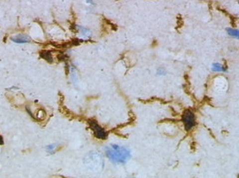
Microglia (green) and neurons (red) living in harmony…for now. Image by Gerry Shaw, EnCor Biotechnology Inc. (Wikicommons).

Alzheimer’s disease (AD) is the most common form of dementia. It’s a neurodegenerative condition characterized by ongoing cognitive decline, loss of functions such as memory, and behavioural abnormalities. AD usually occurs amongst the elderly, and its prevalence is now so high, that its estimated overall cost to society is 5 times that of cancer, heart disease and stroke. While AD was first identified over a century ago, research into its causes has only really begun to gather momentum over the past 30 years. Some think that damage may stem from the formation of toxic ‘plaques’ and ‘tangles’ that appear to be associated with brain cell death. Unfortunately, understanding the triggers of neurodegeneration has become a much more troublesome challenge.

Some teams have begun to look at the contribution of inflammation to the development of brain atrophy (shrinkage). Inflammation is the first response of the immune system to infection/injury. You can recognise inflammation when you graze your skin or sprain an ankle: as well as pain, there may be redness, swelling and the area may feel hot to the touch. This is thanks to the extra blood carrying immune cells to the site of injury to prevent infection and aid repair. The inflammatory response is an innate and usually protective reaction to injury or infection and requires the co-operation between local, ‘resident’ immune cells at the site of injury and circulating immune cells in the bloodstream.

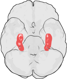
The hippocampi (red) as seen from below the human brain, looking up. (Image from Wikicommons).

The main resident immune cells of the brain are called microglia; they respond to infection and injury to trigger an inflammatory response. Microglia are hyper-sensitive to changes in their local environment. When the brain is injured, they become ‘activated’. They change shape and behave differently, releasing different chemicals which can be toxic to brain cells. Some research has suggested that it’s possible that activated microglia break down the connections between cells in the memory centre of the brain, the hippocampus. Intriguingly, hippocampal damage and memory loss are the primary symptoms of AD.

Several lines of evidence have implicated microglial inflammation in AD, including the observation of activated microglia in the brains with Alzheimer’s, and the possibility that anti-inflammatory drugs may be neuroprotective. However, clinical trials have failed to show any efficacy yet.

Professor Hugh Perry and his research group at the University of Southampton investigate how inflammation contributes to outcome of brain diseases. About ten years ago Perry and his team developed an animal model (the prion mouse) to better understand the complex role of inflammation in AD. A chemical that causes neurodegeneration was injected into the mouse hippocampus (the memory centre of the brain), and the researchers studied the evolution of the resulting prion disease, which bears some similarities to AD. Thirteen weeks after the injection, although the mice appeared normal, there were more activated microglia found in the hippocampus, even compared to surrounding areas of the brain. The researchers then claimed that this microglial activation was pathological, since the mice showed some behavioural disturbances and deficits in learning tasks.

Perry and his team suggested that systemic infections in patients with AD could worsen cell death in the brain, speeding up neuron deterioration and memory loss. (‘Systemic’ infections are so-named because they infect a number of organs and tissues or affect the body ‘system’ as a whole, instead of being localized in one area.)

Resting (top) and activated (bottom) rat microglia after a brain injury. Images by Grzegorz Wicher (Wikicommons).

To look into this idea, researchers looked at differences between the prion mouse with an ‘infection’ (or rather, an injection of a toxin released by bacteria to mimic an infection) or without infection. The prion mouse given a fake infection had twice as many dead brain cells as the uninfected prion mouse. Researchers concluded that the microglia are primed by the ongoing prion disease and so, when the infection is added, they overreact. They then drive the production of a number of inflammatory chemicals, which triggers a whole host of damaging effects on brain cells.

Perry and his team collaborated with other research groups to identify whether the evidence they had gathered would be relevant to AD patients. In a small pilot study of 85 AD patients with moderate cognitive impairment scores over 2 months, they found that those who had infections showed a more cognitive decline than the other patients in the study. This was the first evidence in a clinical setting that systemic infection may affect neurological disease progression. The next study involved 300 AD patients, 50% of whom had a systemic infection within the recorded 6 months. The researchers saw that patients that got an infection within the 6 months suffered three times the rate of cognitive decline, compared to a small cognitive decline in those who had not had an infection.

These fascinating studies have provided the first clinical evidence that as well as inflammation in the brain driving damage, infection and inflammation in the body can also worsen and speed up neurodegeneration. It appears that brain-resident microglia become primed for activation, so that when patients suffer from a bodily infection, their brain cells become more vulnerable to the damaging effects of an inflammatory response.

Not only did this research provide ideas to potentially help AD patients today, but it also formed the basis for an important direction for current disease research. The evidence on the highly complex interplay between the diseased brain and systemic inflammation can be applied, not just to AD, but as a generic concept to many nervous system diseases.

Post by Isabelle Abbey-Vital