Pretty pictures and popular neuroscience go hand-in-hand. People love to see the contours of their brain on an MRI and journalists are drawn to a brain flashing away with activity. There have been some fantastic images from neuroscience in 2013. Here are my favourites, one for each month starting way back in January 2013.
Disclaimer – We own no rights to any of the images on this page. All images are credited to the original authors and copyright holders. The MRC’s Biomedical Picture of the Day has been used as inspiration for some of the images.
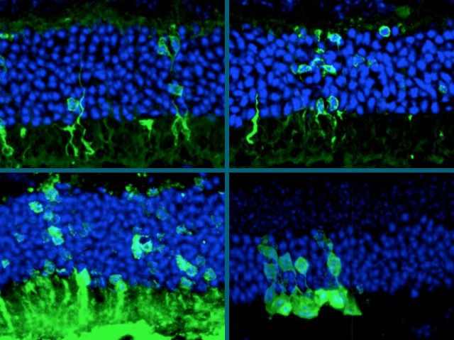
January – New eyes for blindness
Blindness is a major challenge to the neuroscience field. Untreatable blindness is often caused by a degeneration of the light-sensitive cells of the retina. Here, researchers from University College London, UK have injected new photoreceptor cells into the retina of mice with retinal degeneration restoring normal responses to light!
February – Pathfinding connections in the brain
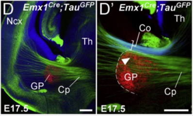
This year there has been a burst of activity in the ‘connectomics’ field. Mapping the connections of the brain is the next big challenge of neuroscience and the main topic of the Human Brain Project in Europe and the BRAIN initiative in the US.
Here, researchers from the École Normale Supérieure in Paris, France looked at how neurons find their way from the thalamus in the middle of the brain to the outermost folds of the cortex.
March – Whole brain activity
http://www.youtube.com/watch?v=KE9mVEimQVU
Seemingly a burning campfire, this is actually a brain flashing with activity. In one of the most impressive images of neuroscience in 2013, researchers from Howard Hughes Medical Institute’s Janelia Farm campus in the US used calcium imaging (see July) to view the activity of a whole brain.
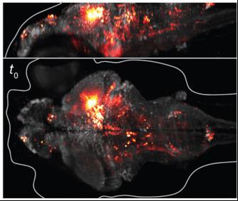
One of the biggest challenges of neuroscience is working out how everything links up together. The most accurate measurements we currently have can only take into account a handful of cells at once. The brilliance of this technique, utilising see-through zebrafish larvae, is that they were able to image more than 80% of the neurons of the brain at once. This can tell you how large populations of cells interact, allowing different regions to work together.
For more info see this article by Mo Costandi in the Guardian.
April – See-through brains
Another amazing technical feat designed to view how the brain links together, in April we were brought CLARITY. By dissolving the opaque fat of a brain whilst keeping the structure intact, researchers led by Karl Deisseroth at Stanford University, California were able to image a whole mouse brain.
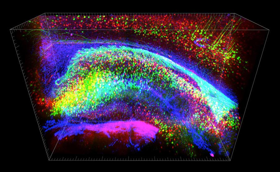
The images from this technique are truly breath-taking. Using this technology, researchers could look in detail at the structure of the brain, giving valuable information of the wiring of different regions. They even imaged part of a post-mortem human brain from an autistic patient, finding evidence of structural defects normally associated with Down’s syndrome.
For more info, see this article in New Scientist.
May – Brainbow 3.0
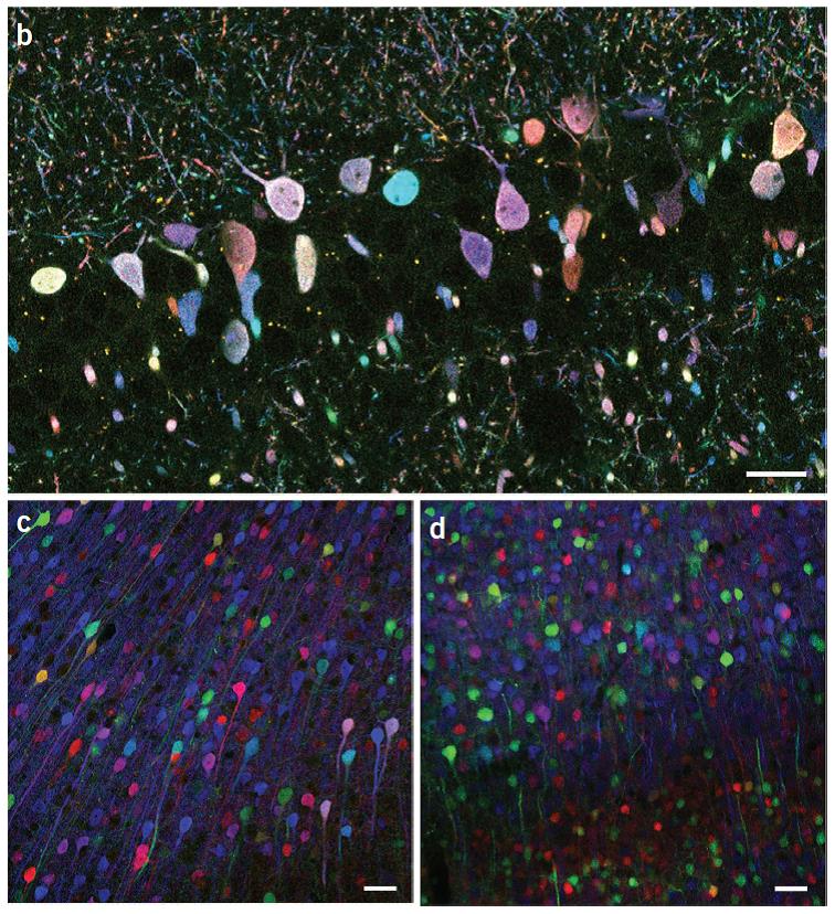
‘Brainbow’ is a transgenic system designed to label different types of cells in many different colours. Prime material for pretty pictures. Take a look at these:
June – Controlling a helicopter with your mind
In June, researchers from the University of Minnesota, USA showed that one could fly a helicopter with their mind! Watch below as the subject guides a helicopter using an EEG skullcap.
July – Better calcium sensors
More calcium imaging now. Calcium imaging works by engineering chemicals that will fluoresce when they encounter calcium. When nerve cells are active, millions of calcium ions flow into the cell at once, therefore a flash of fluorescent light can be seen. Here, researchers from Howard Hughes Medical Institute’s Janelia Farm campus in the US have been working on better, more sensitive calcium sensors. Using these you can colour code neurons based on what they respond to.
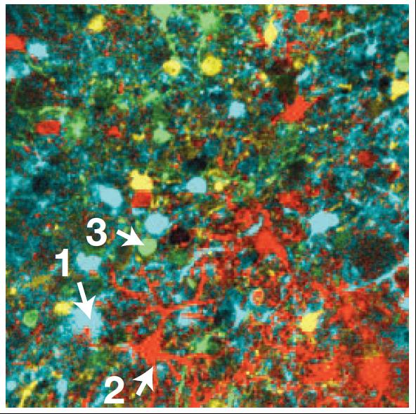
They were also able to record a video of the electrical activity in dendritic spines, the tiny arborisations of nerve cells – see here.
August – Using electron microscopy to connect the brain
Drosophila are wonderful little flies with nervous systems simple enough to get your head around, but complicated enough to be applicable to our own.
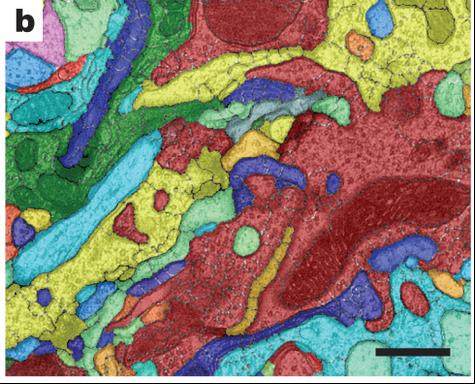
Here, researchers from Janelia Farm (again!) have performed electron microscopy on drosophila brains to connect up neurons across multiple sections. An algorithm colour codes them to line up the same neuron in different sections in what looks like a work by Picasso.
September – Astrocytes to the rescue
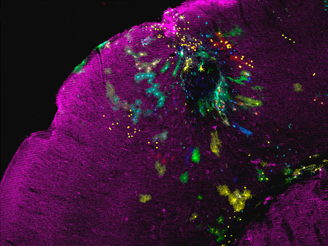
Astrocytes are support cells in the brain which become highly active following brain injury. Here, researchers from Instituto Cajal, CSIC in Madrid, Spain were interested in the different characteristics astrocytes take on when a brain is injured. The injury site can be seen as a dark sphere. Astrocytes with different characteristics have been stained in different colours. For example, the turquoise-coloured astrocytes can be seen forming a protective net around the injury site.
October – Preserved human skulls
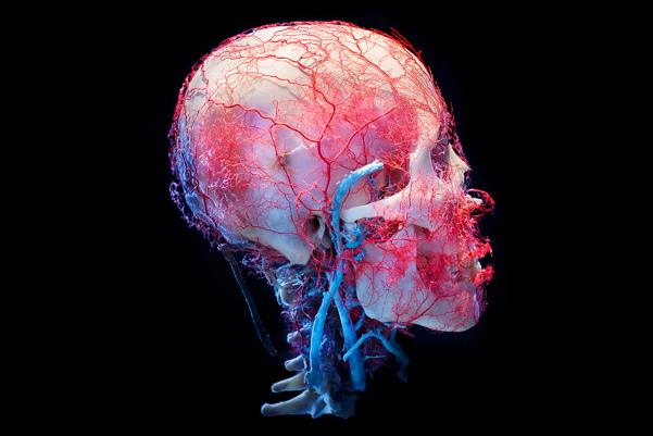
Not strictly neuroscience but these images need to be included. Published in October 2013, The New Cruelty (commissioned by True Entertainment), photographed a series of preserved human skulls.
November – Brain Computing
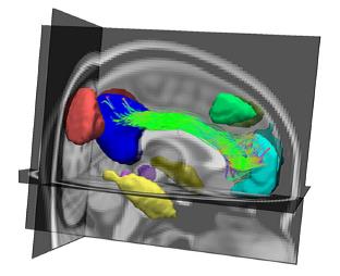
Part of the vision of the Human Brain Project and the BRAIN initiative is to marry anatomy of the brain with computer models to try to produce a working computer model of the brain. This image represents BrainCAT, a software designed to integrate information from different types of brain scan to gain added information about the functionality of the brain.
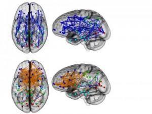
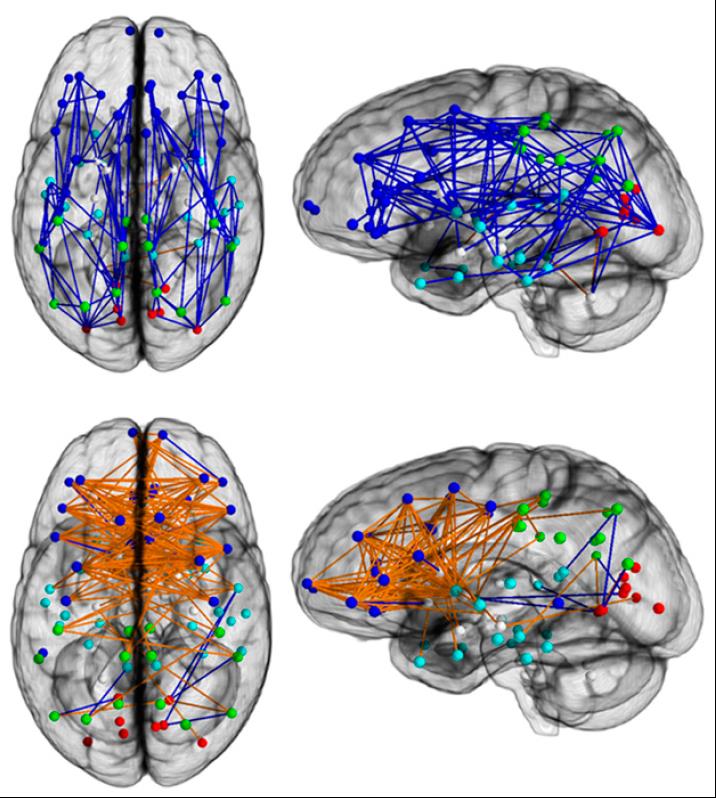
December – Men Are from Mars, Women Are from Venus.
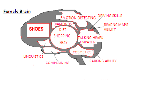
The last month of the year gave us preposterous headlines of ‘proof’ that “Men and women’s brains are ‘wired differently’”. This finally proved why women are from Venus and men are from Mars; why men ‘are better at map reading’ and women are more ‘emotionally intelligent’…. These exaggerated headlines have been kept in check recently on this blog but there’s no denying that the research paper did show some lovely images of male and female brain connections.
So that’s it. 2013 was a year of flashing brains, dodgy connections and overegged hype. Let’s hope there’s even more to come in 2014.
Post by Oliver Freeman @ojfreeman
