PKMZeta: a name to remember.

Will it ever be possible to delete certain painful memories from our conscious brains, as suggested in the film Eternal Sunshine of the Spotless Mind?

We all know what it feels like to remember something, like your first kiss or childhood home, but where in your brain are these memories stored, how do we gain access to them and is it possible to enhance or remove them?

These are questions neuroscientists have spent many years researching. The search for a physical manifestation of memory has taken us on a journey from the truly bizarre (for example a now disproved theory assumed that specific memory molecules existed in the brain and that these could be transfered from one individual to another by eating brain tissue), to our current view that memories are spread throughout the brain and develop when small changes occur in the structure of and connections between neurons (for more detail on synaptic remodeling and plasticity see my previous post). We hope that the more we understand about memory formation and storage, the closer we will come to being able to manipulate them and potentially offer relief to people with memory related illnesses.

PKMZeta structure.

When a memory is first formed a number of proteins become active within participating neurons. These proteins help reshape the neurons thus making the memory permanent. Once this reshaping is complete the proteins involved in the process once again become inactive. It was believed that once reshaping had taken place it would be difficult for us to further modify these neurons to remove or enhance specific memories. However, research conducted within the past 20 years is now questioning this assumption. Researchers have uncovered a protein (PKMZeta) which, unlike others involved in memory formation, remains active in cells long after the initial memory forming event…perhaps indefinitely. This discovery led scientists to question whether PKMZeta may hold the key to maintaining memory and, if so, whether this system could be experimentally manipulated.

Amazingly it seems that this is indeed the case. Scientists have found that blocking the activity of PKMZeta days or even months after learning has taken place can interfere with a rats ability to remember a location, a specific taste or an unpleasant experience. Not only does blocking its activity lead to forgetting, but boosting its activity also has the ability to enhance old faded memories.

Total brainwashing is certainly something we should avoid.

Although the discovery of PKMZeta may be a step forward in finding a treatment for memory disorders, it is important that we proceed with caution and ensure we understand the effects this protein has on the memory system before speculating over its pharmacological value. From current research, scientists believe that the memory enhancing or eradicating effects of PKMZeta are not specific to single memories, indeed they may influence multiple memories at once. Therefore, it is important we understand what memory traces are altered by this protein and how it could be made more selective before considering its wider uses. Removing or enhancing multiple memories non-selectively is certainly not desirable! However, the stage is now set for progress in this field and as our understanding grows there may come a time when we can play a more active role in memory formation and retention.

Post by: Sarah Fox
Your plastic brain

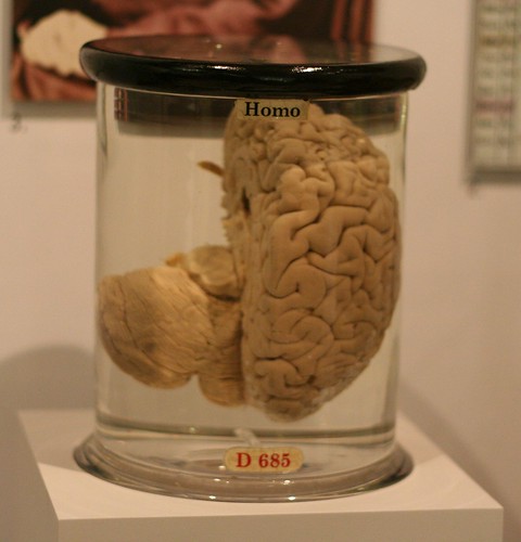
As part of my undergraduate degree I spent a number of sessions studying the anatomical details of preserved human brains. I’m not sure why, but part of me naively expected there to be something awe-inspiringly beautiful about the organ which makes us who we are. Indeed, I can’t deny I was slightly disappointed when our class was presented with a small wrinkled object, dull pink in colour and with a consistency I would liken to children’s play putty. However, looking back on this experience I feel humbled by the knowledge that we are who we are because of this reasonably mundane organ.

Preserved human brain

The human brain weighs around three pounds (a similar weight to three loaves of bread) and houses as many as 100 billion brain cells (neurons). These cells form a number of intricate networks, specialised towards converting sensory information, smell, taste, touch, vision into conscious experience and action.

During our early development neurons forge set communication pathways, the course of which are specified by our genetic code. This genetic framework ensures that we all have the same basic neuronal scaffolding i.e. sensory cells of the eye contact visual areas of the brain whilst those from the ear contact auditory centres etc. (note when things go wrong: Synesthesia). This means that to a large extent we all experience the world in a similar manner. However, the numerous idiosyncrasies which make us individual are likely to depend on the brain’s ability to subtly modify the wiring in many of these networks. Therefore, although some important connections in the brain are hard wired from early childhood, others are referred to as ‘plastic’ and can change throughout life. This ‘plasticity’ underlies the brain’s ability to adapt to new situations and learn new information.

Our brains are plastic…well not literally.

The wiring patterns of cells within brain networks can be altered through three different methods:

  1. The strength of pre-existing neuronal connections may change (also known as synaptic plasticity).
  2. The way individual neurons respond when activated may alter.
  3. New connections may be formed or old ones removed.

Of the above list method 1 is the most widely studied and also happens to be the form of memory my research focuses on. This type of memory requires changes to occur at the region where two neurons connect (commonly referred to as a synapse). Therefore, to fully understand how this process works it is first necessary to appreciate how neurons communicate through these synapses.

A typical neuron can be separated into three functional segments; the dendrites, the axon and the cell body. Although the shape and size of these segments varies from cell to cell, their functions remain largely conserved. Dendrites are the neurons primary input structures, they spread out like branches (hence the name dendrite: from Greek, dendron, ‘tree’) and form synapses with axon terminals of neighbouring cells. Conversely, axons are the neurons primary output structure, transmitting messages from the neuron cell body to other local cells. The cell body, or soma, sits between these two segments supervising the resulting conversation, listening to input from the dendrites and deciding upon the best answer to send via the axon.


Neurons

As the name ‘synaptic plasticity’ indicates, the all-important structure for this form of memory is the synapse. Therefore, using the magnifying glass pictured above, we can now zoom in even further and take a look at how a single synapse on the above neuron functions.


A synapse

The synapse is where an axon terminal from one neuron meets a dendrite of another. Scientists used to think that these two structures touched, however, we now know that a small gap of around 12-20 nano meters (one nano meter is a billionth of a meter) exists between them, this gap is called the ‘synaptic cleft’. The axon terminal communicates by releasing chemicals, known as neurotransmitters, into the cleft. This chemical message crosses the gap and is detected by structures on the adjoining dendrite, thereby notifying the receiving neuron that a communication event has occurred (Note: there are many types of neurotransmitter each of which have different effect on the receiving dendrite).

Individual neurons receive a large number of synaptic connections. These synapses are not all equal, some will have a strong effect on the receiving neuron, encouraging it to send out a message of its own, whilst others will be weaker only exerting a small effect. Indeed, a number of these weak synapses may need to work together to cause the receiving cell to react in the same way as it did to input from one strong synapse. However, the strength of a synapse is not set in stone. Synapses themselves are ‘plastic’ and, given the correct circumstances, can be strengthened or weakened.

Such changes enable us to adapt to new situations, form new memories and are ultimately essential for us to lead a normal life. Hence why it is so important that we understand how and why some synaptic connections become strengthened whilst others do not. This knowledge will not only lead to a better understanding of human memory but will also help us understand what happens when things go wrong, for example during diseases such as Alzheimer’s.

Post by: Sarah Fox


Seahorse and friends: a stroll down memory lane.

I think it’s fitting to begin my first official ‘factual’ post with a fun filled, informal yet informative, trip through my own area of research.

I study a region of the brain known as the hippocampus. This area takes its name from the Latin for seahorse (from ancient Greek: hippos, “horse” and kampos, “sea monster”). The picture below shows a human hippocampus removed from its surrounding brain structures and nicely illustrates where this name comes from:

                                (Source Professor Laszlo Seress, University of Pecs.)

Our hippocampi are located bilaterally (one on either side of the brain) nestled on the underside of the temporal lobe. The image below shows the hippocampus in blue (looking decidedly less like a seahorse) sitting within the temporal lobe, shown in red.

If you were to take the hippocampus pictured above and cut it in half you would find that the inside is not homogeneous, in fact it consists of a number of interlocking cellular semicircles arranged in a circuit (looking rather similar to a swiss roll).

Cells within these curves receive information from a number of brain regions, including visual, olfactory (smell) and auditory (hearing) systems. The cellular circuits found within the hippocampus are believed to be vital for combining such information together to form memories. Indeed, individuals who lack a functioning hippocampus often suffer from a specific inability to form new memories (anterograde amnesia), as illustrated in the films below.

At this point I have the desire to be insanely pedantic, and it’s my blog so I will be! In both these movies the characters are said to have lost their short term memory however, this is not technically correct. Although there is significant debate over the precise functions of the hippocampus, the memory loss experienced by these patients is more likely to represent an inability to transfer new experiences into a long term store rather than representing damage to a short term specific memory system. Or to be significantly less pedantic we can always just say that their swiss roll seahorse is on the frits!

Interestingly damage to the hippocampus does not interfere with all forms of memory. Indeed, individuals with hippocampal damage retain the capacity to learn new skills and often express a sense of familiarity towards recently observed objects, although without any conscious awareness of these memories. For example the, now famous, patient HM (Henry Gustav Molaison) who suffered severe damage to both his left and right hippocampi, retained the capacity to learn new skills such as mirror writing. However, whenever HM was asked to conduct a mirror writing task he argued that he was unable to do so and was always shocked upon discovering his proficiency.
Try it yourself: Print out the star pattern below and hold a small mirror next to this, now (looking only in the mirror) attempt to trace around the star remaining within the two outer lines – the task is much harder than you might think!


8 Sided Star Craft Pattern

Therefore it is believed that the hippocampus is only part of a much larger memory system. Indeed, there are a number of structures situated around the hippocampus within the medial temporal lobe which may all be crucial for different types of memory. I.e. emotional/ fear memory has been linked to the amygdala (a fun link for auditory learners out there ^_^) also object recognition may require the perirhinal cortex. However, the hippocampus is still ultimately recognised as being central to the process of linking many separate aspects of an experience together to form a full flavored memory.

My specific line of research explores how individual cells within the hippocampus form long lasting memories. This basically involves me being a ‘fly on the wall’ listening in on conversations between these cells and understanding how different external factors can influence their communication. This is important for understanding not only how and why we form certain memories but also what happens when things go wrong, for example during debilitating diseases such as Alzheimer’s.

Post by: Sarah Fox